IBS Center for NanoMedicine strives to develop breakthroughs in nanomedicine to gain better understandings and control of nano-bio interface and the phenomena therein. We are developing nanomaterials and nanotools with unique functionality which can lead to new findings in biology and provide the novel approaches in current medicine.
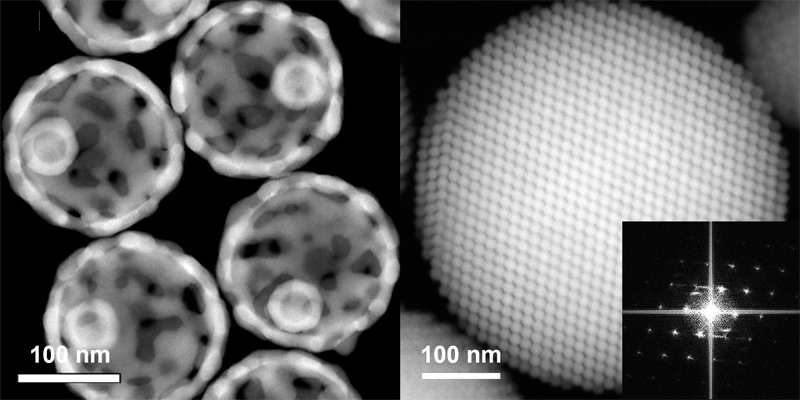
Transmission electron microscope (TEM) images of various types of nanomaterials
We develop programmable nanomaterials with design principles of adaptable and optimized properties. Evolutionary adaptation is a strategy where the nanomaterials response actively under surrounding conditions. Differences in temperature, pH, electromagnetic field, and/or biomolecules can be the factors of adaptations to stimulate the chemical reactions of nanomaterials. The active processes of atomic rearrangements can occur for the morphogenesis of nanomaterials. Such evolutionary nanomaterials exhibit novel physical and chemical properties for further applications. We are currently pursuing to understand the formation and the transformation principles of various types of magnetic, semiconducting, plasmonic, and 2D nanomaterials (Moon et al. Nano Lett. 2017; J. Am. Chem. Soc. 2018).
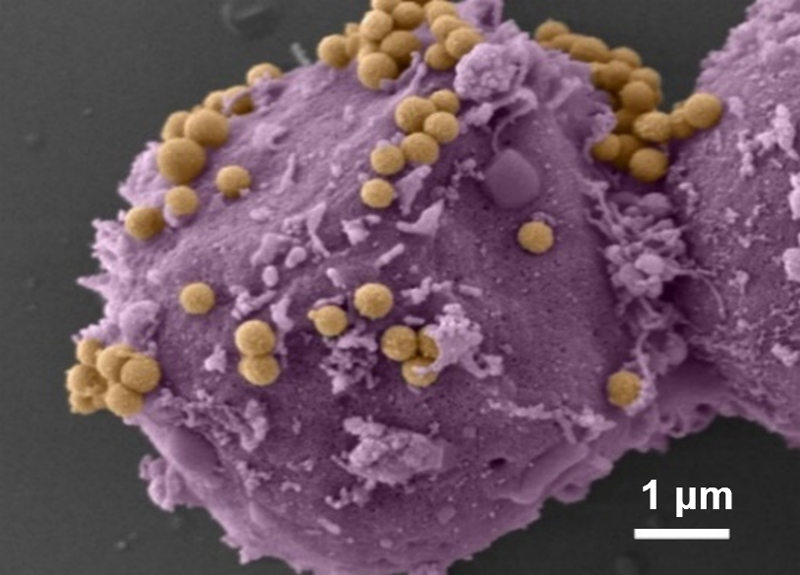
An auditory hair cell with magnetic nanoparticles and an electromagnetic device
Nanoparticle-enabled electronic and mechanical devices are essential for cell signaling control. For example, we are developing magnetic nanoparticle probes as a transducer of magnetic field into a mechanical force for regulating mechanoreceptors of hair cells. These probes can generate time resolved magnetic field and actuate the hair bundles of auditory system to study their oscillatory behaviors (Kim et al. Nano Lett. 2016).
Nano Imaging Reporters
i) Super-resolution MRI
Nanoparticle contrast agents for super-resolution MRIof micro-vessels
Super-resolution MRI contrast agent successfully visualizes the fine structures of vessels in a mouse brain with 100-micrometer spatial resolution. Such nanoparticle contrast agent provides breakthroughs in angiography and tumor imaging. Also, these nanoparticles are capable of identifying characteristics of the biological targets with Magnetic Resonance Tuning (MRET) principle. For example, MRET nanoparticle can accurately detect the level of matrix metalloproteinase-2 (MMP-2) secreted from the tumor to determine whether the tumor is metastatic or not (Shin et al. Nat. Protoc. 2018; Choi et al. Nat. Mater. 2017).
ii) Nano optical 3D imaging of tissue
An optically transparent mouse brain tissue to visualize individual neuronal connectivities in multiscale
We develop a new tissue transformation technology called SHIELD to image thick intact tissues at cell-by-cell resolution in multiscale. The whole process is involved with the development of new tissue preservation method by polyfunctional epoxy, followed by tissue clearing for optical transparentizing. SHIELD allows preservation of tissue archietecture, fluorescence, multiple biomolcules including proteins and nucleic acids simultaneously, which was not possible by other methods previously. As a result, various cell types in organ-scale tissues can be mapped in single-cell resolution by interogating viral fluorescence labels, proteins, RNAs simultaenously. Tracing neuronal projections throughout the intact brain sample can be performed for the construction of true single-cell level neuronal connectivity map. (Park et al. Nat. Biotechnol. 2019).
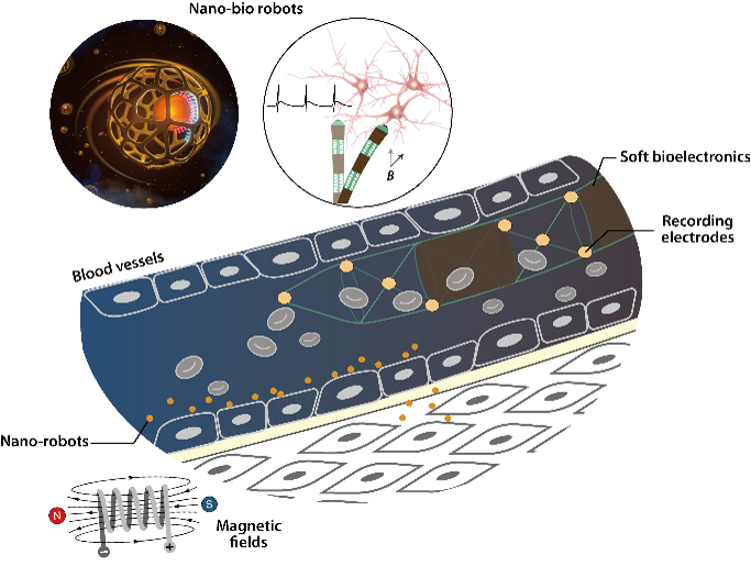
Flexible, transparent, 3D soft nanobio robotics for single-cell monitoring
Our soft nanobio robotics is designed to interface nanodevices with biological cells and organs for digital healthcare. Remote, ultra-flexible, autonomous, and programmable capabilities are key factors of soft-nano bio robotics, which comprises of various types of nanomaterials and biomolecules. These soft nanobio robotic devices will revolutionize the conventional medicine through ultra-sensitive monitoring and manipulations of cells with high precision (Jang et al. Nano Lett. 2019).
Cell function control in single cell and gene level
Nanomachines to control cell fate
The precision nanomedicine we develop utilizes nanotechnology to deliver the future of personalized diagnosis and therapeutics platforms in single cell and gene levels.
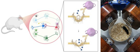
Our single cell nanomachine utilizes nanoscale inputs to regulate biological functions such as gene transcriptions and protein expressions. For instance, magnetic nanoparticles are used to generate mechanical force. Then, mechanoreceptors are pulled to activate cell signaling pathways in a timely and spatially controlled manner, and eventually to direct cellular fates of proliferation, apoptosis, and differentiation (Cho et al. Nat. Mater. 2012; Cho et al. Nano Lett. 2016).